

The approach

What is Minimally Invasive Surgery (MIS)?

Minimally Invasive Techniques By Dr. Nitin Khanna. — Outpatient Surgery, short stay and Faster recovery

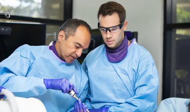
Two clinicians in surgical gowns and gloves at a hands-on training station, one guiding a powered instrument while the other watches
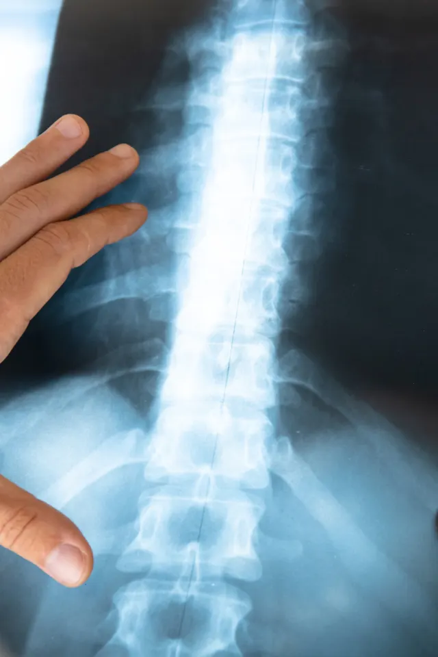
A hand holding a lumbar spine X-ray film up to the light

01 — Conditions

What MIS Treats

In general, the goal of minimally invasive spine (MIS) surgery is to stabilize the vertebral bones and spinal joints and/or relieve pressure being applied to the spinal nerves — often a result of conditions such as:

Conditions MIS is used for

  • Degenerative disc disease
  • Herniated disc
  • Narrowing of the Spinal Canal (Stenosis)
  • Instability of the Spine (spondylolisthesis)
  • Lower Vertebrae Defect (Spondylolysis)
  • Fractured Vertebrae

02 — Benefits & risks

Benefits and Risks

Potential benefits

As opposed to open spine surgery, minimally invasive surgical approaches can be faster, safer, less painful and require less recovery time. Because of the reduced trauma to the muscles and soft tissues (compared to open procedures), the potential benefits are:

  • Better cosmetic results from smaller incisions
  • Less blood loss
  • Reduced risk of muscle and tissue damage (with less or no cutting of the muscle is required)
  • Reduced risk of infection and postoperative pain
  • Many patients can go home the day of surgery, while some will stay in the hospital only overnight
  • Diminished reliance on pain medication post-op

Risks

As with any surgical procedures, no matter how minimal, there are certain risks associated such as:

  • Possible adverse reaction to anesthetic
  • Unexpected blood loss during the procedure
  • Localized infections, no matter how small the incision area
Two clinicians in white coats holding stethoscopes out towards the camera
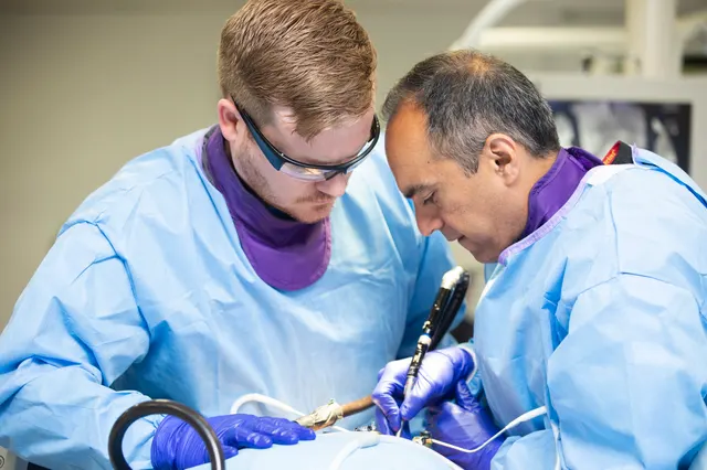
Two clinicians in surgical gowns and gloves work closely together over an instrument at a training station.

03 — How it works

How Minimally Invasive Spine Surgery Works

Minimally invasive surgery of the spine is focused on correcting damage and disorders of the spine (neck and back). Along with lowering the risk of complications, minimally invasive techniques cause less harm to surrounding muscles, bone and other tissue. Often this means less post-operative pain and a faster recovery timeframe.

Traditional

Open surgery

Traditional spine surgery is referred to as open surgery. The procedure utilizes a long incision, down the back with muscles and soft tissue around the spine needing to be moved. In some extreme cases, tissue needs to be removed altogether.

Our approach

Minimally invasive surgery

Our minimally invasive surgical techniques allow us to make a small midline incision and split rather than cut through muscle. This process gently pushes aside muscle and tissue surround the area of focus. Additionally, an intra-operative microscope and x-ray imaging allows the surgeon to use specialized instruments to perform surgery only on the affected area. These factors contribute to decreased post-operative pain and a more rapid return to normal daily activities.

Source: American Association of Neurological Surgeons

Dr. Nitin Khanna

Dr. Khanna's perspective

Not all conditions can be treated with minimally invasive methods and in some cases a traditional "open" procedure is still required. However, I believe in implementing the least invasive approach and prescribing the most advanced forms of treatment wherever possible. Contrary to popular belief, lasers are very rarely used in MIS surgeries. Several methods can be used to minimize trauma during MIS surgery and specific techniques have been deployed for use during minimally invasive procedures. This is a continuously evolving field in which we are driven to stay on the cutting edge.

Nitin Khanna, MD

A clinician points to a lumbar spine X-ray while discussing it with an older patient in a clinic room

04 — Procedures

MIS Procedures

Common Procedures For Conditions of The Neck and Back

DiscectomyVideo

Procedure 01 of 09

Discectomy

Spinal discs are essentially elastic rings with soft material inside that serve as cushions between the vertebral bones. If that elastic ring becomes weakened, the soft tissue inside can extrude — or herniate — outside of the elastic ring. The herniated disc material can compress the nerves passing by, thus causing pain.

A discectomy is the surgical removal of that material pressing on a nerve root or the spinal cord due to a herniated or bulging disc. A discectomy is used to treat a pinched nerve (nerve root compression), bone spur, sciatica, or radiating pain through the limbs called radiculopathy.

If surgical treatment is recommended to trim or remove the herniated disc, it may be possible to perform this procedure with MIS surgery using tubular dilators and, in a minimally invasive discectomy, a special retractor and a microscope or endoscope are used to perform the removal of the herniated disc.

This allows for a smaller incision and a faster recovery. Surgery is completed under general anesthesia, typically taking less than an hour to complete. Most patients go home several hours after the procedure.


Watch the Discectomy video
Spinal Decompression

Procedure 02 of 09

Spinal Decompression

Spinal stenosis, which is a narrowing of the vertebral canal, is a common condition that can result in compression of the nerves. This can produce a variety of symptoms, including pain, numbness and muscle weakness. If surgery is recommended, it may be possible to remove the bone and soft tissues causing the nerve compression through an MIS approach using tubular dilators and a microscope or endoscope. The more common decompressive procedures include laminectomy and foraminotomy.

Anterior Cervical (Neck) Discectomy and Fusion (ACDF)Video

Procedure 03 of 09

Anterior Cervical (Neck) Discectomy and Fusion (ACDF)

Anterior Cervical Discectomy and Fusion (ACDF) is a surgical procedure where the disc is removed through the front of the neck (anterior approach) in order to relieve pressure from the spine (decompress) and relieve pain. This procedure is combined with a fusion surgery in order to stabilize the neck. It is often used to treat herniated disc and degenerative disc disease.

Watch the Anterior Cervical (Neck) Discectomy and Fusion (ACDF) video
Foraminotomy

Procedure 04 of 09

Foraminotomy

A foraminotomy is a procedure that widens the tunnel (foramen) in your back where nerve roots leave your spinal canal. It is used to relieve the symptoms of nerve root compression, also known as a “pinched nerve” or “radiculopathy.” These symptoms may include pain, numbness, tingling or muscle weakness. The procedure can also be performed using a minimally invasive approach that requires no hospitalization. A foraminotomy of the spine can also be used to treat the following conditions:

  • Foraminal stenosis
  • Herniated disc
  • Pinched nerve (nerve root compression)
  • Bone spur
  • Sciatica
  • Radiculopathy
LaminectomyVideo

Procedure 05 of 09

Laminectomy

In a laminectomy, the surgeon removes the lamina (the back part of the vertebra that covers and protects your spinal canal). The procedure takes pressure off the spinal cord or a spinal nerve in cases such as a lumbar stenosis, a herniated disc or bone spur. This procedure can be performed at any level of the spine, using minimally invasive techniques. Patients with single-level or two-level stenosis of the lumbar spine are usually sent home on the day of surgery. A laminectomy can be used to treat spinal stenosis, degenerative disc disease or a herniated disc. Click here to view a video.

Watch the Laminectomy video
Lumbar Spinal FusionVideo

Procedure 06 of 09

Lumbar Spinal Fusion

Spinal fusion is a surgery performed to permanently join two or more bones in the spine so there is no movement between them. To fuse the bones together, the surgeon uses a graft. There are several different types of graft material. For example, an autograft is composed of strips of bone obtained from the area of surgery or another part of your body, such as your pelvic bone, a cadaver bone obtained from a bone bank or made from synthetic material. To promote proper bony fusion, the vertebrae is fixed together with rods, screws, plates or cages to keep the bones from moving until the grafts fully heal. This is called spinal instrumentation. And while fusion surgery does take away some spinal flexibility, in most spinal fusions only small segments of the spine are involved, which do not limit motion much.

Fusion surgery of the spine can be approached from the front, the side or the back. Recent technological advances have allowed fusion surgery to be performed using minimally invasive techniques in select cases. We routinely perform minimally invasive fusion of the lumbar spine from the side (XLIF) as well as from the back (TLIF or PLIF surgery). These less invasive techniques reduce infection rates, minimize blood loss and result in faster patient recovery.

Spinal fusion may relieve symptoms of many back conditions, including the following:

  • Degenerative disk disease
  • Spondylolisthesis
  • Spinal stenosis
  • Scoliosis
  • Fracture
  • Infection
  • Tumor
Watch the Lumbar Spinal Fusion video
Resection of Synovial Cyst

Procedure 07 of 09

Resection of Synovial Cyst

During this procedure, a surgeon removes the synovial cyst and any structures compressing the nerves. This surgery is performed on an outpatient basis with a minimally invasive technique to avoid the need for fusion surgery in select cases.

Sacroiliac Joint FusionVideo

Procedure 08 of 09

Sacroiliac Joint Fusion

SI joint fusion is performed to encourage bone growth over the sacroiliac joint and create one immobile unit for conditions including sacroiliac joint dysfunction that is a direct result of sacroiliac joint disruption and degenerative sacroiliitis. Dr. Khanna utilizes the iFuse Implant System, which is a minimally invasive surgical option. The system is designed to guide the instruments that prepare the bone and facilitate placement of the titanium implants across the sacroiliac joint. The whole MIS SI joint procedure takes about an hour, and recovery time is significantly less than open surgery.

Watch the Sacroiliac Joint Fusion video
Cervical Disc Replacement with Mobi-C

Procedure 09 of 09

Cervical Disc Replacement with Mobi-C

In a surgery with the Mobi-C Cervical Disc, the unhealthy disc is removed, but instead of a bone spacer or plastic implant along with a plate and screws, a Mobi-C is implanted into the disc space. Where a fusion procedure is intended to eliminate motion at the surgery levels, the goal of a surgery with Mobi-C is to allow motion at those levels. The end result is replacement of the damaged disc with the goal of maintaining neck movement. The Mobi-C fits entirely within the disc space.

Watch procedure videos

Become a patient

Ready for the next step in your spine care?

Book an appointment with Dr. Khanna — or send your MRI for a second-opinion review before deciding on surgery.

Recognition